
Electrons are also affected by magnetic fields, and
magnetic focusing had been used for some time in the cathode ray tube. Then in 1926 it was
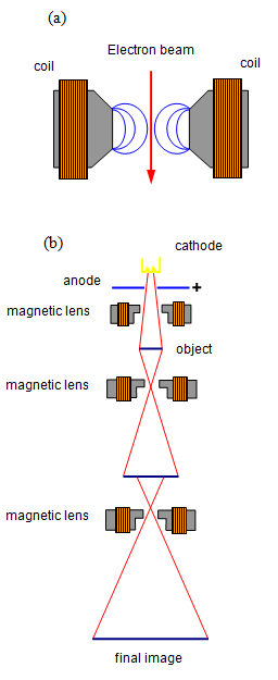
realised that a suitable magnetic field arrangement could be used to act as a magnetic lens,
bringing electrons of a given velocity to a focus and so giving an image of an object.
By
combining several such magnetic lenses a succession of magnifications may be obtained.
shows the similarity between an electron microscope and its optical counterpart. The condenser
lens produces a parallel beam of electrons, which strike the object. Some electrons are
absorbed by the object, some are transmitted and some are scattered sideways. These
scattered electrons cannot pass through the small slit placed in front of the object, and in order
to reduce the number of scattered electrons the object must be very thin.
The
transmitted electrons pass through one or two magnifying lenses and the final image is formed
on a screen or on a photographic plate.
The whole interior of the apparatus must be
maintained at a very high vacuum, otherwise scattering of the electron beam from gas particles
would ruin the image.
The magnification may be varied by altering the current in the
magnetic lenses.
Accelerating voltages of 1 MV have been achieved, and for such an
instrument the field in the lenses reaches a maximum of some 2.5 T using currents of 5A in
3000 turns, giving a focal length of about 5 mm. Such lenses must be cooled and work has
been carried out on superconducting magnets for use in magnetic lenses.
Theoretically the resolution of such an instrument would be very high, but aberration
in the lenses limits it to about 0.1 nm. However magnifications of over 100 000 times are quite
feasible.
Other problems exist at these very high magnifications: the objects that we are
trying to view are little larger, by a factor of 1000 or so, than the electrons in the beam and so
the electron beam distorts the object. Also the very high-energy electrons leak across the
viewing screen, so blurring the image further.