
A ‘normal’ X ray gives only limited information because it is rather like a shadow picture – fine detail within the image may be invisible especially if one organ or bone lies in front of the region of the body being studied.
In the 1970s a method was developed to give much higher quality images including a 3D view of the patient. This method of scanning is called computerised axial tomography (CAT or CT). The name is derived from the Greek for slice (tomos) and read (graphic)
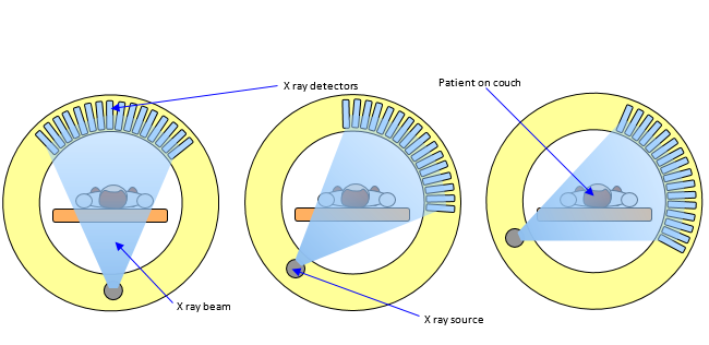
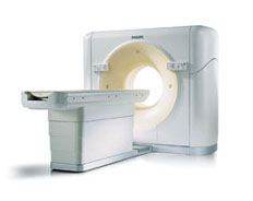
In the CT scanner there is one X ray source but a large number of detectors. The source and the detectors are mounted in a large doughnut shaped machine (see diagram and photo) and the patient is placed inside this on a couch. Each detector records an image and the source and detectors are then rotated around the patient to give views from a variety of direction. The image is called a tomogram. The couch and patient are then moved along the axis of the machine and another set of images is taken. The complete process may take a few minutes or up to half an hour.
This large number of images (many hundreds) are then combined by a computer to give a composite detailed 3D image of the organs under investigation. The development of the CT scanner has been of enormous help in the study of the tumours in cancer patients where images of high quality are essential.
You can compare a ‘normal’ X ray with a CT scan where the X ray is like looking at a book from the outside and the CT scan is like slowly flipping over the pages and reading what is on each of them!
It has been said that the development of the CT scanner was possibly "the greatest legacy" of the Beatles because EMI were able to use some of the massive profits resulting from their record sales to fund scientific research.