
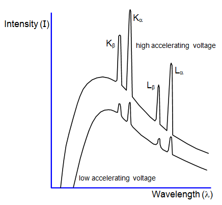
Using X-ray diffraction the spectrum produced by
an X-ray tube can be investigated. Such a spectrum is shown in the diagram, in which the
two curves represent two different accelerating voltages.
The X-ray spectrum can
be considered in two parts:
(a) a continuous background, and (b) a series of sharp
peaks.
The background is due to radiation emitted as the electrons are slowed
down by electromagnetic attraction of the nuclei of the material. The minimum wavelength
and therefore the maximum energy and frequency of an X-ray in this spectrum is produced
when an electron is stopped by just one nucleus. The rest of the curve is produced by
electrons losing only part of their energy during collisions with many nuclei. The minimum
wavelength λm is given by the equation:
where V is the accelerating
voltage and c the velocity of light: the smaller the value of V, the greater the minimum
wavelength will be and the smaller the maximum frequency.
The peaks on the
spectrum are characteristic of the particular target material. Electrons from the inner shells
are removed completely by the bombarding electrons. An electron from an outer energy
level falls back to fill the vacancy emitting a photon of a definite wavelength, thus giving a
sharp peak on the spectrum.